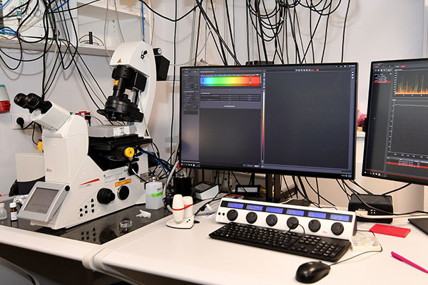
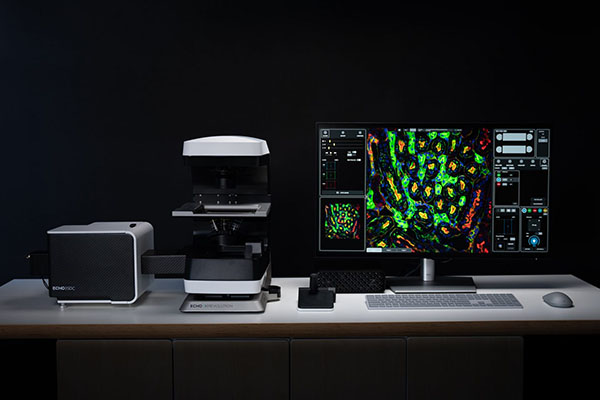
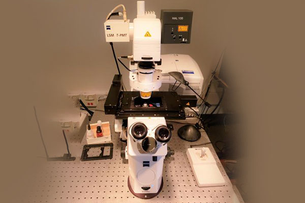
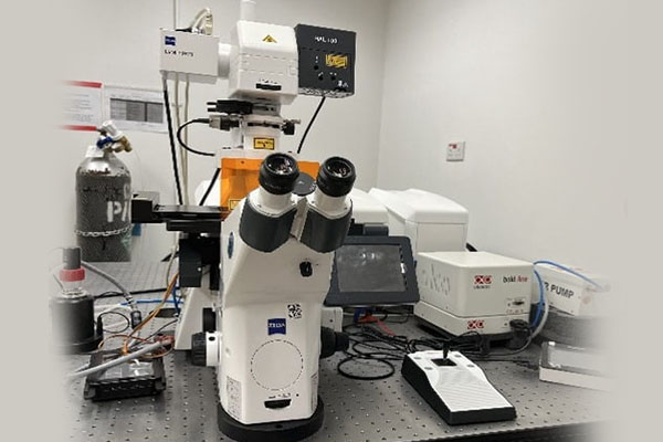
Confocal microscopy
Advantages of Confocal Microscopy in Morphological Studies
The study of morphology—defined as the form, structure, and organization of cells, tissues, and materials—is central to disciplines ranging from biology and medicine to materials science and engineering. While conventional light microscopy opened the door to morphological observation centuries ago, it was limited by optical blurring, poor depth resolution, and an inability to visualize thick specimens in detail. The introduction of confocal microscopy in the 1950s by Marvin Minsky, and its widespread application following the development of laser scanning confocal systems in the 1980s, represented a major technological leap. Confocal microscopy uses a focused laser beam and a pinhole aperture to reject out-of-focus light, enabling high-resolution optical sectioning of specimens. This essay explores the many advantages of confocal microscopy in morphological studies, highlighting its strengths in resolution, optical sectioning, 3D reconstruction, live imaging, quantitative analysis, and interdisciplinary applications.
High-Resolution Imaging
A key advantage of confocal microscopy is its ability to generate high-resolution images of morphological features. Traditional widefield fluorescence microscopes collect light from all focal planes, which creates background blur and reduces clarity. In contrast, confocal systems focus a laser on a specific point in the specimen and collect only the in-focus light through a pinhole, eliminating much of the unwanted background.
This results in sharper, crisper images with excellent contrast. The higher resolution allows scientists to study fine morphological details such as microvilli on epithelial cells, dendritic spines on neurons, or nanostructured coatings on materials. By revealing these minute features, confocal microscopy enhances our ability to understand structure-function relationships at the microscopic level.
Optical Sectioning and Three-Dimensional Reconstruction
Perhaps the most distinctive advantage of confocal microscopy is its capacity for optical sectioning. Instead of physically cutting samples into thin slices, confocal microscopes can capture thin “optical sections” at different focal planes (z-axis). By scanning sequential planes, researchers can reconstruct detailed three-dimensional (3D) models of morphological structures.
This is particularly valuable in biology, where cells and tissues are inherently three-dimensional. For example, the morphology of neuronal networks, tumor spheroids, or embryonic tissues can be studied in their entirety without the artifacts of mechanical sectioning. In materials science, confocal microscopy provides volumetric analysis of coatings, porous structures, and composite layers. The ability to visualize morphology in 3D transforms confocal microscopy into a powerful tool for both qualitative observation and quantitative measurement.
Non-Destructive Imaging of Intact Samples
Another advantage is that confocal microscopy is non-destructive and does not require extensive sample preparation. Unlike transmission electron microscopy (TEM), which necessitates ultra-thin sectioning, confocal microscopy can image intact cells, tissues, and materials. Biological specimens can often be studied in a near-native state, preserving their morphology.
Moreover, confocal microscopy is compatible with live specimens, meaning that dynamic morphological changes can be observed in real time. This is crucial for studies of processes such as cell migration, wound healing, organelle movement, and developmental biology. The ability to study morphology in living systems, without significant disturbance, is a major strength of confocal microscopy.
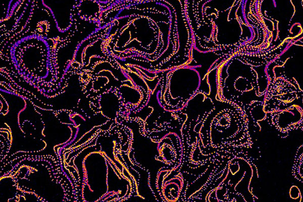
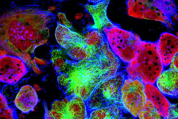
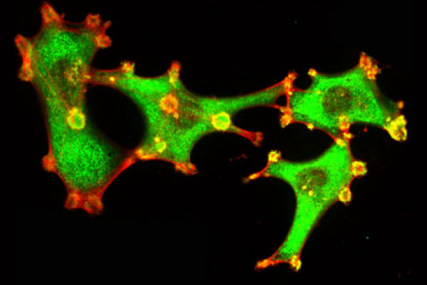
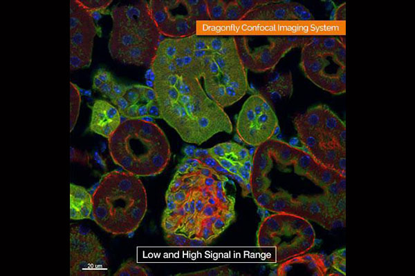
Examples of Confocal Microscopy
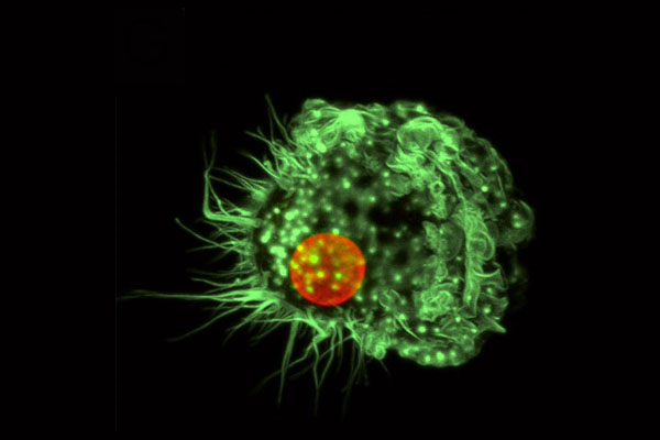

Morphology in Live-Cell Imaging
Confocal microscopy is widely regarded as one of the best techniques for live-cell morphological studies. Its ability to minimize out-of-focus blur ensures that the morphology of cells can be monitored even within thick, multicellular environments. Combined with fluorescent labeling, confocal microscopy allows researchers to track the morphology of specific structures over time, such as the cytoskeleton, cell membranes, or nuclei.
This has been transformative in developmental biology, where confocal imaging has revealed how cells change shape, divide, and interact during embryogenesis. In neuroscience, confocal microscopy has enabled the study of dendritic spine morphology and synapse formation, which are critical for understanding learning and memory. The temporal dimension of live-cell imaging adds an invaluable layer of insight to morphological studies.
Multicolor Imaging and Molecular Specificity
Morphological studies often require distinguishing between different components of a specimen. Confocal microscopy excels in this area by enabling multicolor fluorescence imaging. Multiple fluorescent dyes or proteins can be used simultaneously, each targeting a different structure.
For example, in cell biology, the nucleus can be labeled with DAPI, the actin cytoskeleton with phalloidin, and mitochondria with a specific tracker dye. Confocal microscopy can then visualize these structures in separate channels or merge them into a composite image, providing a holistic view of cellular morphology. In materials research, different phases or layers of a composite material can be selectively labeled and imaged in detail.
This molecular specificity, combined with morphological imaging, allows researchers to not only see shape and structure but also correlate them with function and composition.
Quantitative Morphological Analysis
Confocal microscopy is not limited to qualitative imaging; it also provides opportunities for quantitative morphometric analysis. Using digital reconstruction and image processing software, confocal datasets can be analyzed to measure parameters such as cell size, shape, volume, thickness, and surface topology.
This quantitative aspect is critical in many fields. For instance, in cancer research, confocal imaging can quantify changes in nuclear or cytoskeletal morphology associated with malignancy. In materials science, the thickness of coatings or the porosity of scaffolds can be measured with high precision. By combining imaging with measurement, confocal microscopy provides robust datasets that go beyond visual inspection.
Reduced Background and Enhanced Contrast
Another advantage is the ability of confocal systems to reduce background noise and produce high-contrast images. The pinhole rejects out-of-plane fluorescence, which is particularly beneficial when studying thick specimens. Conventional widefield microscopes often produce blurred images when imaging such samples, but confocal microscopy retains clarity.
This enhanced contrast is crucial for accurately interpreting morphological details. For example, in developmental biology, individual cells can be clearly distinguished within a tissue, while in materials science, surface defects and boundaries can be precisely defined.
Broad Applicability Across Disciplines
The advantages of confocal microscopy in morphology extend across numerous fields:
- Cell Biology: Reveals the architecture of organelles, cytoskeleton, and membranes with 3D clarity.
- Neuroscience: Enables detailed studies of neuronal morphology, synaptic connections, and network formation.
- Developmental Biology: Tracks morphological changes during embryogenesis and tissue morphogenesis.
- Medical Research: Used to study tumor morphology, angiogenesis, and tissue engineering scaffolds.
- Material Science: Characterizes coatings, microstructures, and polymer blends in 3D.
- Plant Biology: Examines cell walls, stomata, and vascular tissue architecture.
The versatility of confocal microscopy in imaging diverse specimens highlights its central role in modern morphological research.
Lorem ipsum dolor sit amet, consectetur adipiscing elit. Ut elit tellus, luctus nec ullamcorper mattis, pulvinar dapibus leo.
Limitations and Contextual Advantages
While confocal microscopy offers many advantages, it is important to acknowledge certain limitations, such as photobleaching of fluorescent dyes, limited penetration depth, and slower imaging compared to widefield methods. Nevertheless, when evaluating advantages in the context of morphology, confocal microscopy stands out as an unmatched tool for non-destructive, high-resolution, and 3D imaging.
Conclusion
Confocal microscopy has transformed the way morphology is studied across the biological and material sciences. Its unique combination of high resolution, optical sectioning, 3D reconstruction, and live imaging capabilities provides insights that are unattainable with conventional microscopy. By offering multicolor imaging, quantitative analysis, and enhanced contrast, confocal microscopy allows researchers to explore not just the static architecture of specimens, but also their dynamic morphological changes in real time.
Ultimately, the greatest advantage of confocal microscopy in morphological studies is its ability to bridge structure and function while preserving specimen integrity. Whether revealing the fine details of a neuron, tracking the growth of a tumor, or analyzing the surface of a biomaterial, confocal microscopy provides a window into morphology that is both precise and holistic. Its contributions continue to advance science and medicine, ensuring its position as a cornerstone technology for morphological research in the 21st century.