TRANSMISSION ELECTRON MICROSCOPY
Advantages of Transmission Electron Microscopy in Morphological Studies
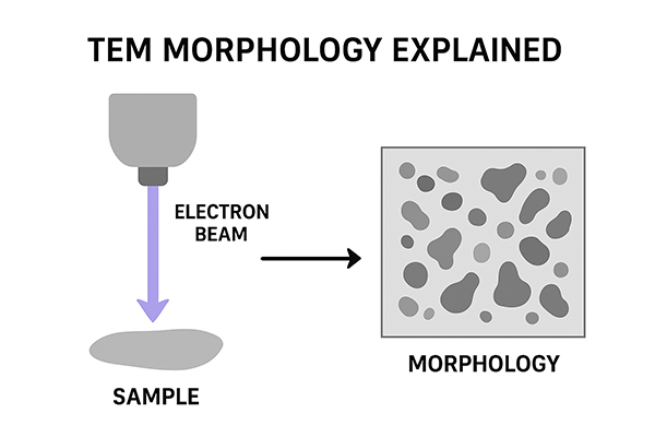
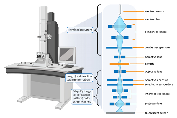
The study of morphology—encompassing the form, structure, and organization of biological and material systems—has long been a central pursuit in science. Traditional light microscopy provided early insights into morphology, but it was constrained by the diffraction limit of visible light, which restricted resolution to approximately 200 nanometers. The invention and subsequent refinement of the Transmission Electron Microscope (TEM) revolutionized morphology studies by enabling visualization at sub-nanometer scales. TEM relies on the transmission of high-energy electrons through ultra-thin specimens, allowing researchers to study internal structures in unprecedented detail. This essay explores the many advantages of TEM in morphological studies, highlighting its unparalleled resolution, ability to reveal internal ultrastructure, versatility, analytical capabilities, and contributions across disciplines.
Unparalleled Resolution and Magnification
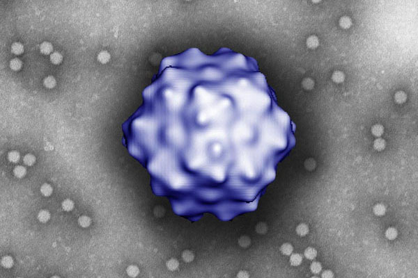
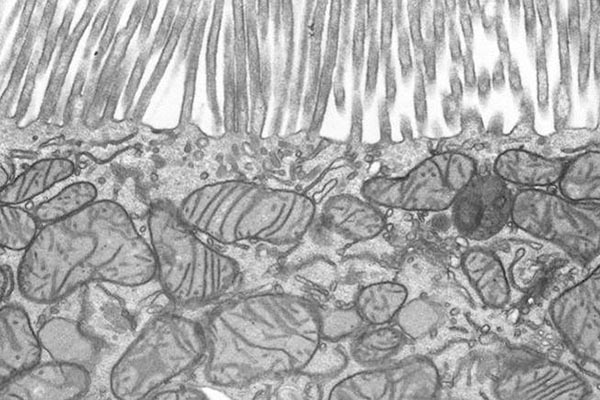
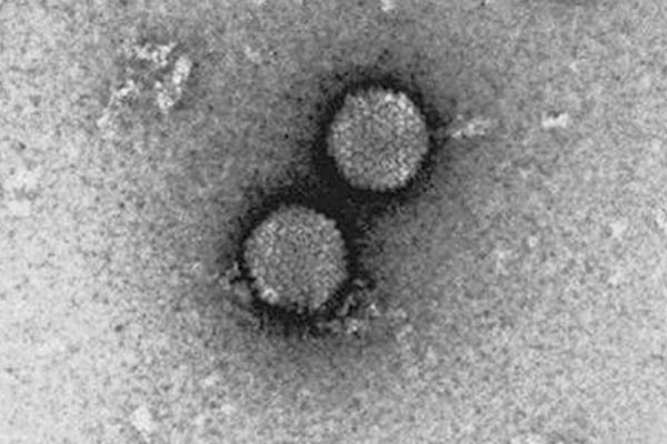
The most celebrated advantage of TEM is its ability to achieve ultra-high resolution. Because electrons have much shorter wavelengths than visible light photons, they can resolve structural details at the atomic level. Modern TEM systems achieve resolutions in the range of 0.1–0.2 nanometers, which is orders of magnitude finer than the limit of optical microscopy. This extraordinary resolution allows TEM to visualize the precise arrangement of atoms in a crystal lattice, the double membrane of mitochondria, or the fine organization of viral capsids.
In terms of magnification, TEM can operate across a broad spectrum, from modest magnifications of a few hundred times to over a million times. This flexibility enables both holistic and highly localized morphological studies within the same specimen. The ability to zoom from organelles to macromolecules, or from grain boundaries to atomic dislocations, makes TEM indispensable for morphology research.
Internal Morphology and Ultrastructure
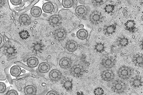
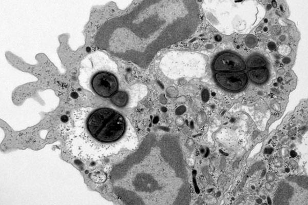
Unlike Scanning Electron Microscopy (SEM), which primarily reveals surface morphology, TEM uniquely provides insights into internal morphology. By transmitting electrons through thin specimens, TEM allows scientists to visualize the arrangement of structures deep inside cells, tissues, or materials. For biological studies, this translates into detailed images of cell organelles such as the nucleus, endoplasmic reticulum, ribosomes, and chloroplasts. Viruses and protein complexes, invisible under light microscopy, become accessible with TEM.
In materials science, TEM reveals the internal structure of nanoparticles, thin films, and crystalline materials. Dislocations, stacking faults, and precipitates within metals or semiconductors become evident under TEM, providing essential information for understanding mechanical strength, conductivity, or catalytic activity. Thus, TEM enables morphological studies that go far beyond surface characterization, reaching into the very core of materials and cells.
Crystallographic and Structural Information
TEM does more than simply image morphology—it also provides crystallographic information. Techniques such as electron diffraction and high-resolution TEM (HRTEM) allow researchers to study crystal symmetry, lattice spacing, and orientation with atomic precision. This is particularly valuable in nanomaterials research, where morphology and crystallinity are closely linked to functionality.
For instance, in semiconductor studies, TEM reveals grain boundaries and point defects that affect electronic properties. In catalysis, the morphology of nanoparticles, along with their crystal facets, determines catalytic efficiency. In metallurgy, TEM elucidates how heat treatment affects the microstructure of alloys. The ability to combine morphological visualization with crystallographic analysis sets TEM apart from most other imaging techniques.
Analytical Versatility
Another advantage of TEM lies in its integration with spectroscopic techniques. Energy-dispersive X-ray spectroscopy (EDS or EDX), electron energy-loss spectroscopy (EELS), and related analytical methods can be coupled with TEM to provide compositional data alongside morphology. This allows researchers to not only see how a sample is structured but also to identify the chemical elements present within specific regions.
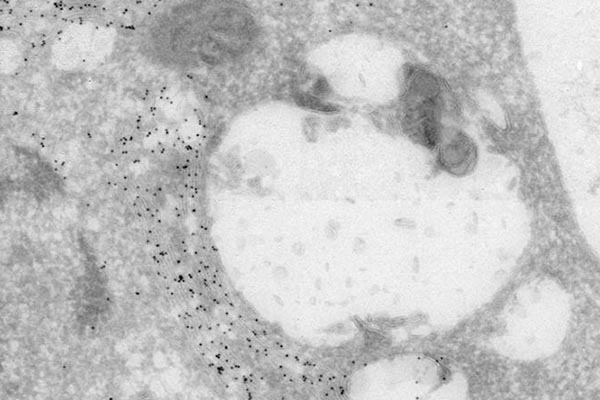
For example, in biological specimens, TEM-EDS can reveal the localization of heavy metal stains or nanoparticles within a cell. In materials science, TEM-EELS can detect variations in oxidation states, bonding environments, or elemental distributions at the nanoscale. This correlative approach—linking morphology with composition—gives researchers a holistic understanding of structure-function relationships.
Examples of Transmission Electron Microscopy
Visualization of Defects and Fine Features
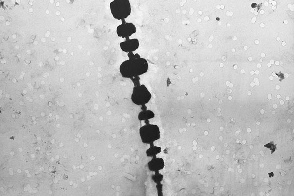
Morphological studies are not only about ideal structures but also about imperfections, as defects often control the behavior of materials and biological systems. TEM excels at detecting structural irregularities. In crystalline materials, TEM reveals dislocations, vacancies, stacking faults, voids, and grain boundaries with extraordinary clarity. These features play critical roles in determining mechanical strength, conductivity, and optical properties.
In polymers and composites, TEM reveals phase separation, fiber distribution, and nanofiller dispersion. In biological systems, it highlights subtle differences in organelle morphology that may correspond to health or disease states. The ability of TEM to detect both normal and abnormal morphology makes it an essential diagnostic and research tool.
Contrast Mechanisms for Morphology
TEM provides multiple contrast mechanisms, each offering unique advantages for morphological studies. Mass-thickness contrast arises because heavier atoms or thicker regions scatter more electrons, appearing darker in images. Diffraction contrast highlights crystalline regions, making grain boundaries and crystallites visible. Phase contrast, particularly in HRTEM, enables direct visualization of atomic arrangements.
These contrast mechanisms enrich morphological analysis by providing information not only about shapes and sizes but also about density, crystallinity, and internal organization. This multi-dimensional insight makes TEM morphology studies much more informative than simple imaging techniques.
Broad Applicability Across Disciplines
TEM’s advantages in morphological studies extend to a wide range of scientific disciplines:
- Biology and Medicine: TEM has revealed the ultrastructure of viruses, bacteria, and organelles, contributing to breakthroughs in cell biology and virology. Pathologists use TEM to diagnose certain diseases by observing characteristic ultrastructural changes.
- Nanotechnology: TEM is indispensable for studying nanoparticle size, shape, crystallinity, and defects. Morphological control at the atomic scale underpins advances in quantum dots, carbon nanotubes, and other nanostructures.
- Materials Science: TEM has transformed metallurgy, ceramics, and polymer science by providing detailed microstructural insights. Fracture studies, heat treatment analysis, and thin film characterization all rely on TEM morphology.
- Chemistry and Catalysis: Morphology often determines catalytic efficiency. TEM helps correlate particle morphology with surface reactivity, supporting the rational design of catalysts.
- Geology and Environmental Science: TEM reveals the morphology and composition of mineral inclusions, aerosols, and nanoclays, shedding light on processes ranging from volcanism to climate dynamics.
Non-Destructive Nanoscale Characterization
Although TEM requires ultra-thin sections, the technique is largely non-destructive at appropriate operating conditions. With careful electron beam management, the same specimen area can be observed repeatedly without significant damage. This enables comparative studies of morphological changes under varying conditions, such as heating, irradiation, or mechanical stress. Modern cryo-TEM techniques also preserve delicate biological structures in near-native states, allowing researchers to study morphology without chemical artifacts.
Limitations and Contextual Advantages
It is important to acknowledge that TEM does have limitations, such as complex sample preparation, the need for ultra-thin sections, and operation in high vacuum. However, when weighed against its advantages, TEM remains the gold standard for studying internal morphology at the nanoscale. Its ability to reveal ultrastructure with atomic resolution cannot be matched by SEM, light microscopy, or most other imaging modalities.
Conclusion
Transmission Electron Microscopy has fundamentally reshaped morphological studies by providing direct access to the nanoscopic and atomic world. Its advantages are manifold: ultra-high resolution, the ability to visualize internal ultrastructure, crystallographic analysis, integration with spectroscopy, detection of defects, and applicability across numerous disciplines. By combining imaging with analytical power, TEM enables researchers to correlate form, structure, and function with extraordinary precision.
The transformative impact of TEM lies not only in its technical strengths but also in its role as a bridge between morphology and function. From elucidating the architecture of viruses to analyzing defects in semiconductors, TEM has empowered scientists to link structural features with performance and behavior. As technology advances, TEM continues to expand its capabilities, incorporating cryogenic methods, tomography, and in-situ experimentation. Ultimately, TEM remains the most powerful and versatile tool for morphological studies, offering unparalleled insights into the hidden structures that define both the living and material worlds.