SCANNING ELECTRON MICROSCOPY
Advantages of Scanning Electron Microscopy in Morphological Studies
The study of morphology, which encompasses the form, structure, and surface characteristics of materials and biological specimens, is central to a wide range of scientific disciplines. From materials science and nanotechnology to biology, medicine, and geology, morphology provides key insights into the relationship between structure and function. The advent of Scanning Electron Microscopy (SEM) has revolutionized the field of morphological studies by offering a unique combination of high-resolution imaging, three-dimensional visualization, and versatile analytical capabilities. Unlike conventional optical microscopy, SEM employs electrons rather than photons to generate images, thereby overcoming the diffraction limit of visible light and revealing fine structural details on the nanometer scale. This essay explores the numerous advantages of SEM in morphological studies, emphasizing its resolution, depth of field, versatility, and impact across diverse fields of research.
High Resolution and Magnification
One of the most significant advantages of SEM lies in its ability to achieve high magnification with exceptional resolution. While optical microscopes are limited to a resolution of about 200 nanometers due to the wavelength of visible light, SEM can achieve resolutions as fine as 1–2 nanometers with modern field emission systems. This allows researchers to observe intricate morphological features that would otherwise remain invisible. For instance, SEM enables the detailed visualization of nanoparticle shape, bacterial surface appendages, or the roughness of fracture surfaces in metals. The ability to magnify from a few tens to hundreds of thousands of times provides both broad overviews and close-up analyses within a single instrument, making SEM a versatile tool for morphological investigation.
Superior Depth of Field
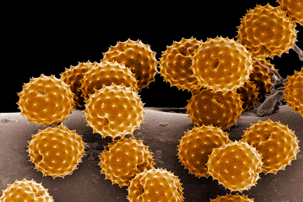
Another key advantage of SEM in morphological studies is its superior depth of field compared to optical microscopy. The depth of field refers to the thickness of the specimen that remains in focus at a given magnification. In optical systems, particularly at higher magnifications, the depth of field becomes extremely shallow, often restricting the observer to a very thin slice of the sample. SEM, on the other hand, provides a much greater depth of field, giving images a distinctive three-dimensional appearance. This is particularly valuable when studying complex surface structures such as porous materials, geological formations, or biological specimens like pollen grains and insect exoskeletons. The pseudo-3D visualization allows researchers to better appreciate the texture, layering, and topology of surfaces.
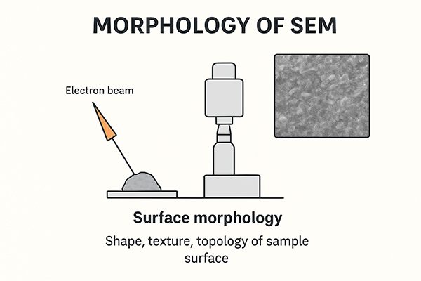
Detailed Surface Morphology
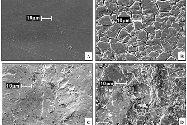
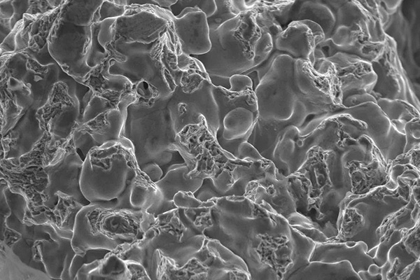
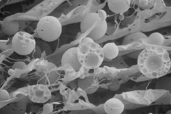
Morphology is often defined by surface features, and SEM is specifically designed to probe surface characteristics with remarkable clarity. The technique relies primarily on secondary electrons emitted from the top few nanometers of the specimen’s surface, which provide fine topographical detail. This makes SEM ideal for observing surface roughness, grain boundaries, cracks, and other microstructural attributes that directly influence material properties. For example, in metallurgy, SEM has been indispensable in fracture analysis, where the morphology of fractured surfaces reveals whether the failure was ductile, brittle, or fatigue-induced. In nanotechnology, SEM allows precise characterization of nanostructures, enabling researchers to control and optimize synthesis processes. In biology, SEM reveals the intricate textures of cellular and extracellular structures, ranging from the microvilli of epithelial cells to the sculpted exteriors of diatoms.
Wide Range of Sample Types
SEM’s versatility in handling diverse specimens is another advantage in morphological studies. Unlike Transmission Electron Microscopy (TEM), which requires ultra-thin sections, SEM can accommodate bulk samples with minimal preparation. Metals, ceramics, polymers, biological tissues, and even archaeological artifacts can be imaged with SEM. While non-conductive samples typically require a conductive coating of gold, platinum, or carbon, this step is relatively straightforward compared to the elaborate preparation needed for TEM. Furthermore, with the development of environmental SEM (ESEM), it is now possible to image hydrated and non-conductive samples without extensive preparation, thereby preserving their natural morphology. This has opened new avenues in biological sciences, where delicate samples can be studied in near-native conditions.
Three-Dimensional Imaging and Intuitive Visualization
The pseudo-3D quality of SEM images gives researchers an intuitive sense of surface morphology that is difficult to achieve with other techniques. Unlike two-dimensional projection images produced by TEM, SEM images mimic the way humans perceive surfaces in everyday life. This enhances interpretability and communication of morphological findings, not only within scientific communities but also in teaching and industrial applications. For example, SEM images of microcracks or corrosion pits provide engineers with immediately understandable evidence of material degradation, while SEM images of microorganisms serve as compelling illustrations in biological education.
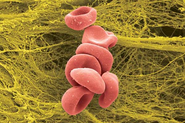
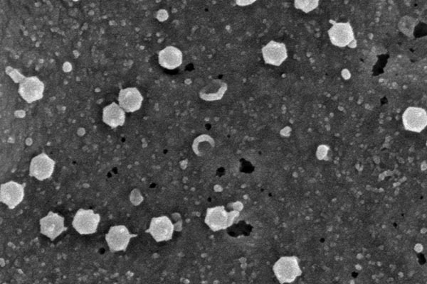
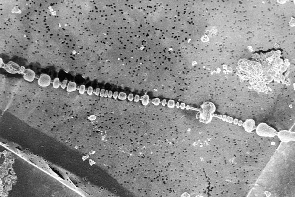
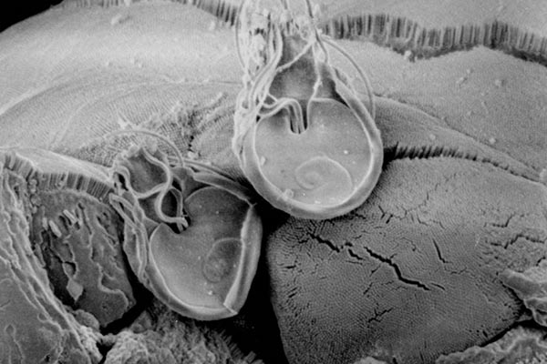
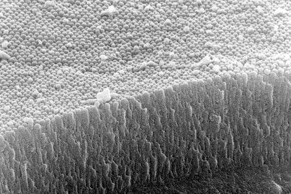
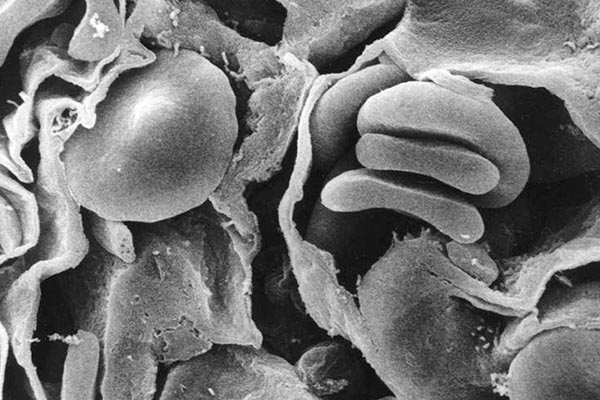
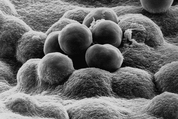
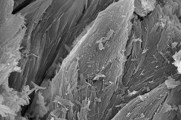
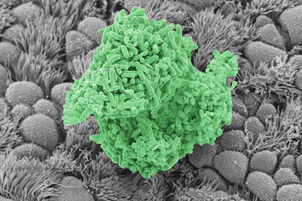
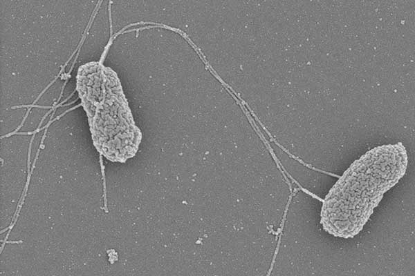
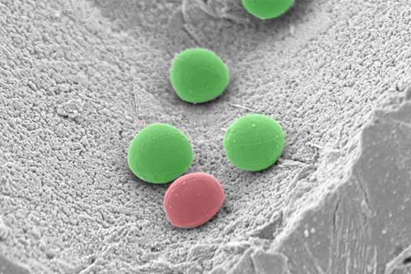
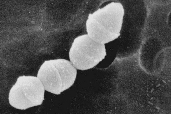
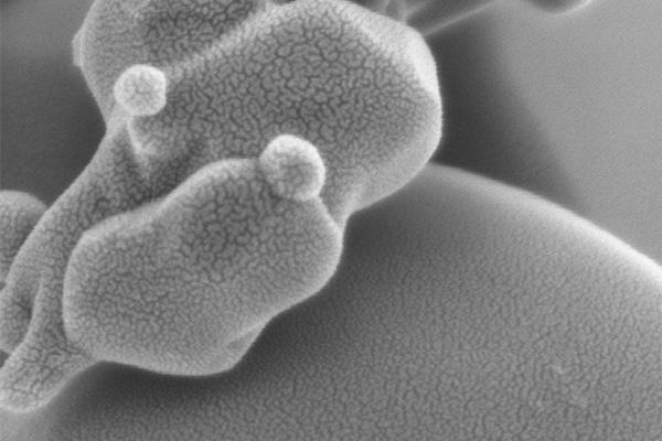
Examples Of Scanning Electron Microscopy
Analytical Capabilities Alongside Morphology
A distinctive advantage of SEM is its ability to combine morphological imaging with compositional analysis. Modern SEM instruments are frequently equipped with detectors such as energy-dispersive X-ray spectroscopy (EDS or EDX), which provide elemental composition of the sample. This integration allows researchers not only to study the surface morphology but also to correlate it with chemical characteristics. In geological studies, for example, SEM coupled with EDS can reveal both the shape and the elemental makeup of mineral grains. In materials science, the correlation between morphology and composition is essential for understanding alloy microstructures, corrosion processes, or the distribution of catalysts in porous supports.
Non-Destructive Characterization
Although SEM does involve interaction between the electron beam and the sample, the technique is generally considered non-destructive, especially at lower accelerating voltages. Samples can often be imaged repeatedly without significant damage, enabling longitudinal studies of morphological changes. For instance, polymers, composites, or biological samples may be studied under different conditions, such as heating or stretching, to observe how morphology evolves in real time. This capability makes SEM particularly valuable in research and industrial quality control, where the integrity of samples must be maintained.
Applications Across Disciplines
The advantages of SEM in morphology extend to a wide range of scientific and industrial fields:
- Materials Science and Engineering: SEM is essential for characterizing microstructures, coatings, and fracture surfaces. Morphological analysis provides clues about mechanical strength, wear resistance, and failure mechanisms.
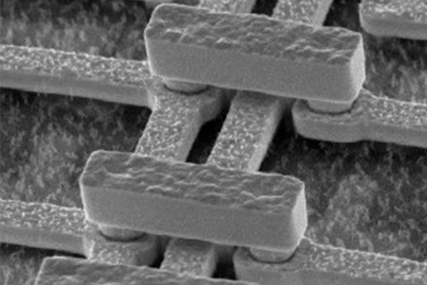
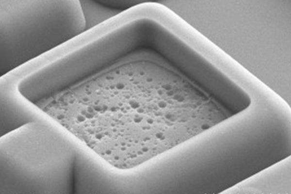
- Nanotechnology: Morphological control at the nanoscale is central to nanomaterials synthesis. SEM provides direct visualization of nanoparticle size, shape, and distribution, enabling optimization of fabrication methods.
- Biology and Medicine: SEM has revealed the surface architecture of cells, tissues, and pathogens, aiding in understanding biological function and disease processes. Morphological studies of bacteria, for example, help in identifying species or studying biofilm formation.
- Geology and Earth Sciences: SEM morphology of rocks, soils, and fossils provides insights into formation history, weathering processes, and paleobiology.
- Forensic Science: SEM morphology of fibers, residues, or gunshot particles supports forensic investigations by linking evidence to sources.
Limitations and Contextual Advantages
While emphasizing the advantages, it is important to acknowledge that SEM is not without limitations. It requires vacuum conditions, conductive coatings for insulating samples, and may induce charging or beam damage in delicate materials. However, compared to other imaging modalities, SEM offers a uniquely advantageous balance of resolution, depth of field, versatility, and compositional analysis. These advantages make it the method of choice for many morphological studies where surface structure plays a central role.
Conclusion
Scanning Electron Microscopy has emerged as an indispensable tool for morphological studies across disciplines. Its advantages stem from a combination of high resolution, superior depth of field, detailed surface imaging, three-dimensional visualization, and integrated analytical capabilities. SEM accommodates a broad spectrum of samples with relatively simple preparation, and with the advent of environmental SEM, it can even handle hydrated and non-conductive specimens in near-native states. By providing rich morphological information that bridges structure and function, SEM not only advances fundamental scientific research but also supports applied sciences, industry, and education. The transformative impact of SEM lies in its ability to reveal the hidden surface world with clarity and depth, offering researchers unprecedented opportunities to explore, understand, and manipulate morphology at micro- and nanoscales.