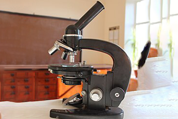
LIGHT MICRSCOPY
Advantages of Light Microscopy in Morphological Studies
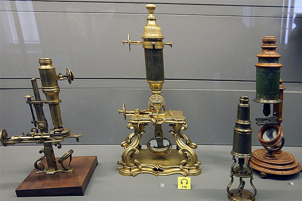
The study of morphology, which involves analyzing the form, structure, and arrangement of cells, tissues, and organisms, has always been central to biology, medicine, and material sciences. Since the 17th century, when pioneers like Robert Hooke and Antonie van Leeuwenhoek first peered into the microscopic world using rudimentary light microscopes, our understanding of biological and structural morphology has expanded immensely. Among the many tools developed for microscopy, light microscopy remains one of the most fundamental and widely used techniques. Despite the emergence of advanced technologies such as electron microscopy and confocal microscopy, light microscopy continues to play a critical role in morphological studies due to its unique combination of accessibility, versatility, and ability to study living specimens.
This essay examines in detail the key advantages of light microscopy in morphological research, highlighting its strengths in direct visualization, simplicity, live-cell imaging, affordability, rapid sample preparation, diversity of techniques, and broad applicability across scientific disciplines.
Direct Visualization of Morphology
One of the foremost advantages of light microscopy is its ability to provide direct visualization of cellular and tissue morphology. Under a light microscope, researchers can easily observe fundamental features such as cell shape, size, organization, and arrangement within tissues. Light microscopy enables the study of natural color in specimens or enhances visibility through staining methods, which highlight structures like nuclei, cytoplasm, and extracellular components.
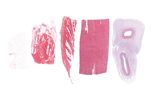
For instance, in histology, thin tissue sections stained with hematoxylin and eosin (H&E) reveal the detailed architecture of organs. In microbiology, Gram staining differentiates bacteria based on their cell wall morphology. These techniques demonstrate the capacity of light microscopy to quickly and effectively display essential morphological details that are critical for diagnosis and research.
Simplicity and Accessibility
Another major advantage is the simplicity and accessibility of light microscopy. Compared to electron microscopes, which require extensive training, vacuum systems, and costly infrastructure, light microscopes are straightforward to operate. They are found in nearly every biology classroom, clinical laboratory, and research institution worldwide.
This accessibility ensures that morphological study is not restricted to highly specialized laboratories but can be carried out by students, clinicians, and researchers with varying levels of expertise. As a result, light microscopy democratizes scientific inquiry, making morphological analysis broadly available to both advanced scientists and beginners.
Observation of Living Specimens
Perhaps one of the most significant advantages of light microscopy over techniques such as transmission electron microscopy (TEM) is its ability to study living cells and organisms in real time. Since light microscopes do not require a vacuum chamber or destructive preparation methods, researchers can place living samples directly under the microscope and monitor dynamic morphological changes as they occur.
This is particularly important in cell biology, where processes such as mitosis, motility, endocytosis, and cell differentiation can be directly observed. For example, the shape changes of white blood cells as they migrate toward pathogens or the elongation of plant root hairs during growth can be followed under light microscopy. Such live-cell imaging provides invaluable insights into both static and dynamic morphology.
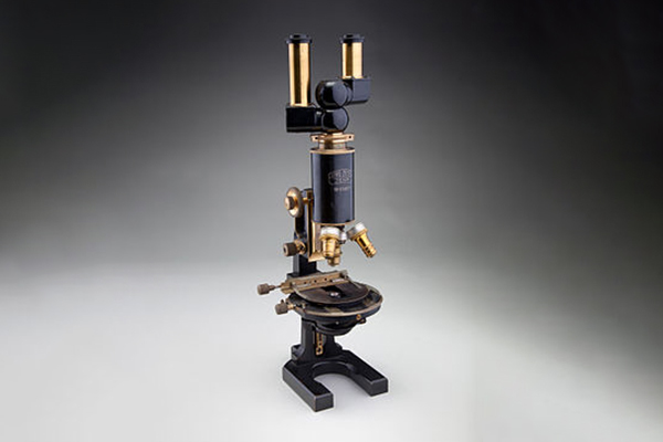
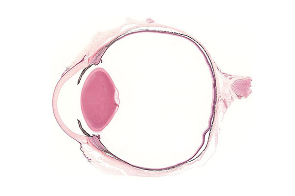
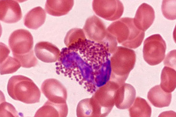
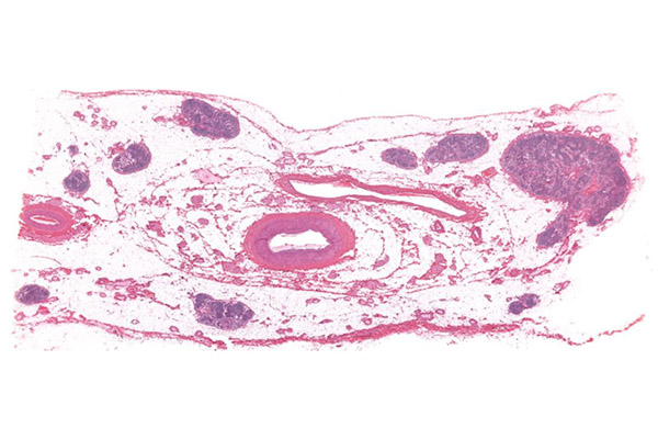
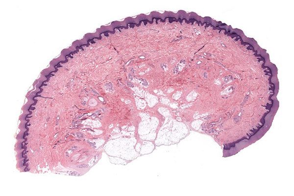
Examples Of Light Microscopy:

Natural Color and Staining Flexibility
Unlike electron microscopy, which produces grayscale images, light microscopy allows the observation of specimens in natural color. This provides an immediate and intuitive understanding of morphology. Furthermore, a wide range of staining techniques can be applied to highlight specific features.
- Histological stains such as H&E differentiate between cellular structures.
- Special stains like Periodic acid–Schiff (PAS) can highlight carbohydrates and glycogen.
- Vital stains can label living cells without killing them, allowing morphological observation alongside physiological function.
These staining techniques not only increase contrast but also expand the scope of morphological studies by correlating form with biochemical composition.
Cost-Effectiveness
Light microscopy is also cost-effective, especially compared to advanced imaging technologies such as confocal or electron microscopy. The initial purchase cost of a standard light microscope is relatively low, and ongoing maintenance expenses are minimal. Consumables such as glass slides, cover slips, and basic stains are inexpensive and widely available.
This affordability allows schools, small clinics, and laboratories in developing regions to access microscopy for morphological studies. It ensures that morphological analysis remains an essential part of education and medical diagnostics worldwide.
Rapid Sample Preparation
Another advantage lies in the simplicity and speed of sample preparation. For light microscopy, preparing a specimen often requires minimal steps such as smearing, sectioning, or applying a stain. In many cases, fresh samples can be observed directly without any treatment.
This contrasts with electron microscopy, where specimens must be fixed, dehydrated, embedded, sectioned ultrathin, and coated with heavy metals—a time-consuming process that also risks introducing artifacts. The rapid preparation associated with light microscopy makes it especially valuable in clinical settings, where quick morphological assessment can aid diagnosis and treatment decisions.
Variety of Techniques for Morphological Studies
Modern light microscopy includes a wide range of specialized techniques that expand its utility for morphological studies:
- Brightfield microscopy: The most common form, used for stained tissues and cells.
- Darkfield microscopy: Enhances contrast by using scattered light, useful for small or transparent structures.
- Phase-contrast microscopy: Allows visualization of unstained, living cells by highlighting differences in refractive index.
- Differential interference contrast (DIC): Produces high-contrast images with a pseudo-3D appearance, ideal for fine morphological details.
- Fluorescence microscopy: Enables visualization of specific structures tagged with fluorescent dyes or proteins.
These diverse approaches allow light microscopy to reveal morphology at multiple levels, from whole tissues to subcellular organelles, and under different physiological states.
Broad Applicability Across Disciplines
The versatility of light microscopy ensures its widespread use across various fields:
- Biology and Medicine: Studying tissue histology, blood smears, microbial morphology, and cellular organization.
- Pathology: Diagnosing cancer and other diseases through morphological examination of biopsy samples.
- Microbiology: Identifying bacterial morphology (cocci, bacilli, spirilla) and staining properties.
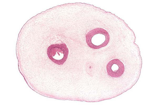
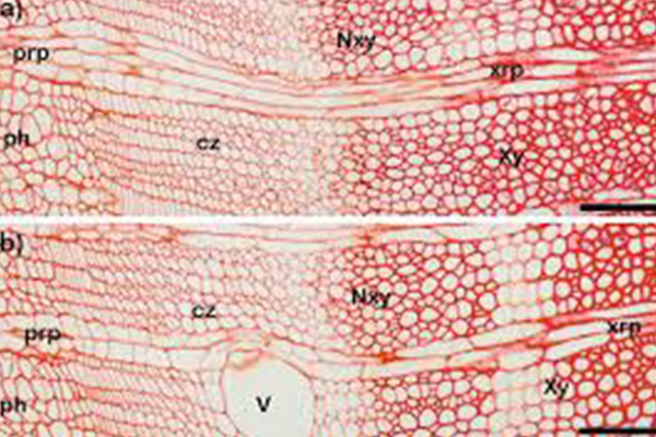
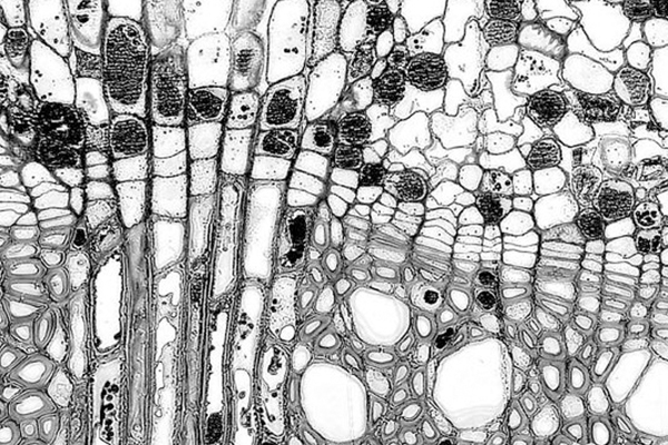
- Botany: Observing plant tissues, stomata, and vascular systems.
- Material Science: Examining fibers, crystals, thin films, and composite materials.
The wide applicability of light microscopy across disciplines underscores its importance as a universal tool for morphological study.
Reduced Risk of Artifacts
Because light microscopy requires relatively simple preparation, there is a lower risk of introducing structural artifacts compared to methods like TEM, where fixation and sectioning may distort the specimen. This ensures that observed morphology is often closer to the natural state of the sample.
Limitations and Contextual Advantages
It is important to acknowledge that light microscopy has limitations, such as lower resolution (limited by the diffraction of light, around 200 nm) and less ability to visualize ultrastructural details compared to TEM or SEM. However, in the context of morphological studies—especially those requiring live imaging, quick preparation, and cost-effective analysis—the advantages of light microscopy are highly significant.
Conclusion
Light microscopy remains one of the most valuable and versatile tools in morphological studies. Its ability to provide direct visualization of cell and tissue architecture, combined with its simplicity, affordability, and capacity to observe living specimens, ensures its continued relevance despite the advent of more advanced imaging technologies. The diversity of techniques such as phase-contrast, fluorescence, and differential interference contrast further enhances its utility in revealing fine morphological details.
Ultimately, the strength of light microscopy lies in its balance of accessibility, practicality, and effectiveness. By enabling scientists, clinicians, and students to explore morphology in both living and preserved specimens, light microscopy continues to serve as the foundation of structural biology and medical diagnostics. Its enduring role highlights not only its historical importance but also its modern relevance as a cornerstone technique in the study of form and structure.